Colposcopy - directed biopsy
Definition
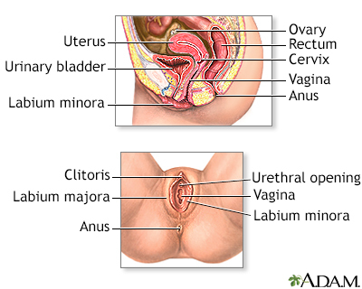
A colposcopy is a special way of looking at the cervix. It uses a light and a low-powered microscope to make the cervix appear much larger. This helps your health care provider find and then biopsy abnormal areas in your cervix.
Alternative Names
Biopsy - colposcopy - directed; Biopsy - cervix - colposcopy; Endocervical curettage; ECC; Cervical punch biopsy; Biopsy - cervical punch; Cervical biopsy; Cervical intraepithelial neoplasia - colposcopy; CIN - colposcopy; Precancerous changes of the cervix - colposcopy; Cervical cancer - colposcopy; Squamous intraepithelial lesion - colposcopy; LSIL - colposcopy; HSIL - colposcopy; Low-grade colposcopy; High-grade colposcopy; Carcinoma in situ - colposcopy; CIS - colposcopy; ASCUS - colposcopy; Atypical glandular cells - colposcopy; AGUS - colposcopy; Atypical squamous cells - colposcopy; Pap smear - colposcopy; HPV - colposcopy; Human papilloma virus - colposcopy; Cervix - colposcopy; Colposcopy
How the Test is Performed
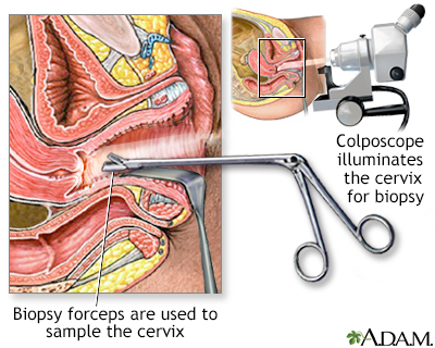
You will lie on a table and place your feet in stirrups, to position your pelvis for exam. The provider will place an instrument (called a speculum) into your vagina to see the cervix clearly.
The cervix and vagina are gently cleaned with a vinegar or iodine solution. This removes the mucus that covers the surface and highlights abnormal areas.
The provider will place the colposcope at the opening of the vagina and examine the area. Photographs may be taken. The colposcope does not touch you.
If any areas look abnormal, a small sample of the tissue will be removed using small biopsy tools. Several samples may be taken. Sometimes a tissue sample from inside the cervix is removed. This is called endocervical curettage (ECC).
How to Prepare for the Test
There is no special preparation. You may be more comfortable if you empty your bladder and bowel before the procedure.
Before the exam:
- Do not douche (this is never recommended).
- Do not place any products into the vagina.
- Do not have sex for 24 hours before the exam.
- Tell your provider if you are pregnant or could be pregnant.
This test should not be done during a heavy period, unless it is abnormal. Keep your appointment if you are:
- At the very end or beginning of your regular period
- Having abnormal bleeding
You may be able to take ibuprofen or acetaminophen (Tylenol) before the colposcopy. Ask your provider if this is OK, and when and how much you should take.
How the Test will Feel
You may have some discomfort when the speculum is placed inside the vagina. It may be more uncomfortable than a regular Pap test.
- Some women feel a slight sting from the cleansing solution.
- You may feel a pinch or cramp each time a tissue sample is taken.
- You may have some cramping or slight bleeding after the biopsy.
- Do not use tampons or put anything in the vagina for several days after a biopsy.
Some women may hold their breath during pelvic procedures because they expect pain. Slow, regular breathing will help you relax and relieve pain. Ask your provider about bringing a support person with you if that will help.
You may have some bleeding after the biopsy, for about 2 days.
- You should not douche, place tampons or creams into the vagina, or have sex for up to a week afterward. Ask your provider how long you should wait.
- You can use sanitary pads.
Why the Test is Performed
Patient Education Video: Cervical cancer
Colposcopy is done to detect cervical cancer and changes that may lead to cervical cancer.
It is most often done when you have had an abnormal Pap smear or HPV test. It may also be recommended if you have bleeding after sexual intercourse.
Colposcopy may also be done when your provider sees abnormal areas on your cervix during a pelvic exam. These may include:
- Any abnormal growth on the cervix, or elsewhere in the vagina
- Genital warts or HPV
- Irritation or inflammation of the cervix (cervicitis)
The colposcopy may be used to keep track of HPV, and to look for abnormal changes that can come back after treatment.
Normal Results
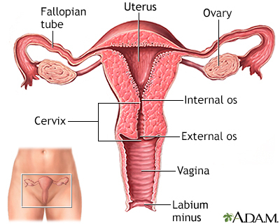
A smooth, pink surface of the cervix is normal.
A specialist called a pathologist will examine the tissue sample from the cervical biopsy and send a report to your doctor. Biopsy results most often take 1 to 2 weeks. A normal result means there is no cancer and no abnormal changes were seen.
What Abnormal Results Mean
Your provider should be able to tell you if anything abnormal was seen during the test, including:
- Abnormal patterns in the blood vessels
- Areas that are swollen, worn away, or wasted away (atrophic)
- Cervical polyps
- Genital warts
- Whitish patches on the cervix
Abnormal biopsy results may be due to changes that can lead to cervical cancer. These changes are called dysplasia, or cervical intraepithelial neoplasia (CIN).
- CIN I is mild dysplasia
- CIN II is moderate dysplasia
- CIN III is severe dysplasia or very early cervical cancer called carcinoma in situ
Abnormal biopsy results may be due to:
- Cervical cancer
- Cervical intraepithelial neoplasia (CIN) -- precancerous tissue changes that are also called cervical dysplasia
- Cervical warts (infection with human papilloma virus, or HPV)
If the biopsy does not determine the cause of abnormal results, you may need a procedure called a cold knife cone biopsy.
Risks
After the biopsy, you may have some bleeding for up to a week. You may have mild cramping, your vagina may feel sore, and you may have a dark discharge for 1 to 3 days.
A colposcopy and biopsy will not make it more difficult for you to become pregnant, or cause problems during pregnancy.
Contact your provider right away if:
- Bleeding is very heavy or lasts for longer than 2 weeks.
- You have pain in your belly or in the pelvic area.
- You notice any signs of infection (fever, foul odor, or discharge).
Gallery
References
Cohn DE, Ramaswamy B, Christian B, Bixel K. Malignancy and pregnancy. In: Resnik R, Lockwood CJ, Moore TR, Greene MF, Copel JA, Silver RM, eds. Creasy and Resnik's Maternal-Fetal Medicine: Principles and Practice. 8th ed. Philadelphia, PA: Elsevier; 2019:chap 56.
Khan MJ, Werner CL, Darragh TM, et al. ASCCP colposcopy standards: role of colposcopy, benefits, potential harms and terminology for colposcopic practice. Journal of Lower Genital Tract Disease. 2017;21(4):223-229. PMID: 28953110 pubmed.ncbi.nlm.nih.gov/28953110/.
Newkirk GR. Colposcopic examination. In: Fowler GC, ed. Pfenninger and Fowler's Procedures for Primary Care. 4th ed. Philadelphia, PA: Elsevier; 2020:chap 124.
Salcedo MP, Phoolcharoen N, Schmeler KM. Intraepithelial neoplasia of the lower genital tract (cervix, vagina, vulva): etiology, screening, diagnosis, management. In: Gershenson DM, Lentz GM, Valea FA, Lobo RA, eds. Comprehensive Gynecology. 8th ed. Philadelphia, PA: Elsevier; 2022:chap 29.
