Eye floaters
Alternative Names
Specks in your vision
Information
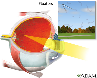
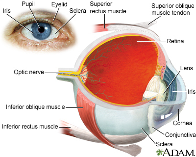
The floating specks you sometimes see in front of your eyes are not on the surface of your eyes, but inside them. These floaters are bits of cell debris that drift around in the fluid (vitreous) that fills the back of your eye. They may look like spots, specks, bubbles, threads, or clumps. Most adults have at least a few floaters. There are times when they may be more visible than at other times, such as when you are reading.
Most of the time floaters are harmless. However, they can be a symptom of a tear in the retina. (The retina is the layer in the back of the eye.) If you notice a sudden increase in floaters or if you see floaters along with flashes of light in your side vision, this may be a symptom of a retinal tear or detachment. Go to an eye doctor or emergency room if you have these symptoms.
Sometimes a dense or dark floater will interfere with reading.
Recently, a laser treatment has been developed that may be able to break up this type of floater so that it is not so bothersome.
Gallery
References
Broadhead GK, Hong T, Chang AA. To treat or not to treat: management options for symptomatic vitreous floaters. Asia Pac J Ophthalmol (Phila). 2020;9(2):96-103. PMID: 32097127 pubmed.ncbi.nlm.nih.gov/32097127/.
Crouch ER, Crouch ER, Grant TR. Ophthalmology. In: Rakel RE, Rakel DP, eds. Textbook of Family Medicine. 9th ed. Philadelphia, PA: Elsevier Saunders; 2016:chap 17.
Guluma K, Lee JE. Ophthalmology. In: Walls RM, Hockberger RS, Gausche-Hill M, eds. Rosen's Emergency Medicine: Concepts and Clinical Practice. 9th ed. Philadelphia, PA: Elsevier; 2018:chap 62.
Shah CP, Heier JS. YAG laser vitreolysis vs sham YAG vitreolysis for symptomatic vitreous floaters: a randomized clinical trial. JAMA Ophthalmol. 2017;135(9): 918-923. PMID: 28727887 pubmed.ncbi.nlm.nih.gov/28727887/.
