Eye - foreign object in
Alternative Names
Foreign body; Particle in the eye
Information
The eye will often flush out small objects, like eyelashes and sand, through blinking and tearing. DO NOT rub the eye if there is something in it. Wash your hands before examining the eye.
Patient Education Video: What to do when something gets in your eye
Examine the eye in a well-lit area. To find the object, look up and down, then from side to side.
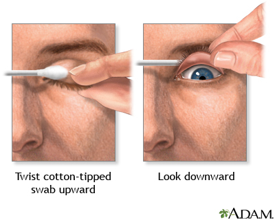
- If you can't find the object, it may be on the inside of one of the eyelids. To look inside the lower lid, first look up then grasp the lower eyelid and gently pull down. To look inside the upper lid, you can place a cotton-tipped swab on the outside of the upper lid and gently fold the lid over the cotton swab. This is easier to do if you are looking down.
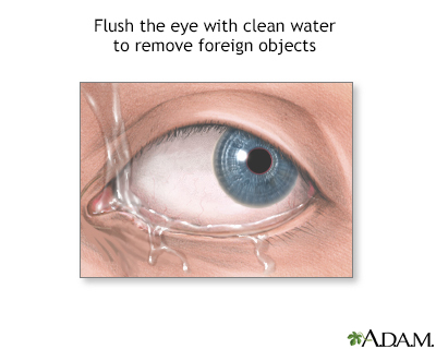
- If the object is on an eyelid, try to gently flush it out with water or eye drops. If that does not work, try touching a second cotton-tipped swab to the object to remove it.
- If the object is on the white of the eye, try gently rinsing the eye with water or eye drops. Or, you can GENTLY touch a cotton swap to the object to try to remove it. If the object is on the colored part of the eye, DO NOT attempt to remove it. Your eye may still feel scratchy or uncomfortable after removing an eyelash or other tiny object. This should go away within a day or two. If you continue to have discomfort or blurred vision, get medical help.
Contact your health care provider and DO NOT treat yourself if:
- You have a lot of eye pain or sensitivity to light.
- Your vision is decreased.
- You have red or painful eyes.
- You have flaking, discharge, or a sore on your eye or eyelid.
- You have had trauma to your eye, or you have a bulging eye or a drooping eyelid.
- Your dry eyes do not get better with self-care measures within a few days.
If you have been hammering, grinding, or could have come in contact with metal fragments, DO NOT attempt any removal. Go to the nearest emergency room immediately.
Gallery
References
Crouch ER, Crouch ER, Grant TR. Ophthalmology. In: Rakel RE, Rakel DP, eds. Textbook of Family Medicine. 9th ed. Philadelphia, PA: Elsevier; 2016:chap 17.
Fowler GC. Corneal abrasions and removal of corneal or conjunctival foreign bodies. In: Fowler GC, ed. Pfenninger and Fowler's Procedures for Primary Care. 4th ed. Philadelphia, PA: Elsevier; 2020:chap 200.
Knoop KJ, Dennis WR. Ophthalmologic procedures. In: Roberts JR, Custalow CB, Thomsen TW, eds. Roberts and Hedges' Clinical Procedures in Emergency Medicine and Acute Care. 7th ed. Philadelphia, PA: Elsevier; 2019:chap 62.
Thomas SH, Goodloe JM. Foreign bodies. In: Walls RM, Hockberger RS, Gausche-Hill M, eds. Rosen's Emergency Medicine: Concepts and Clinical Practice. 9th ed. Philadelphia, PA: Elsevier; 2018:chap 53.