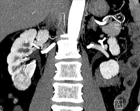
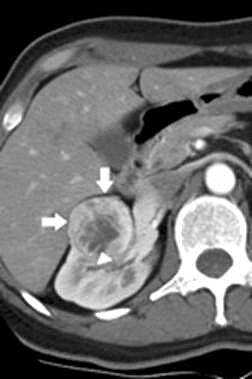
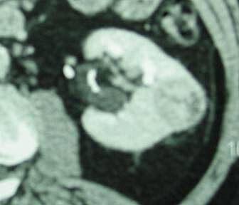
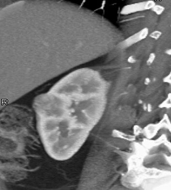
For patients diagnosed with a small (i.e. < 4 cm) kidney tumor (4 Figures below), laparoscopic and robotic partial nephrectomy provides patients with a safe and minimally invasive technique for removal of the tumor, while preserving the remainder of the healthy kidney.
By removing only the tumor and not the entire kidney, patients have a significantly lower risk of kidney failure down the road. These minimally invasive approaches have the advantage of improved cosmesis, reduced pain, blood loss, and hospital stay, as compared to conventional open surgery with similar cure rates. In select patients with larger tumors, partial nephrectomy may not be feasible and therefore radical (i.e. complete) nephrectomy may be required.
To learn more about kidney (or renal cell) cancer, please click here.
The Surgery
Laparoscopic and robotic partial nephrectomy requires that patients undergo a general anesthesia. While operative time varies from one individual to another, the average operating time is approximately 3-4 hours.
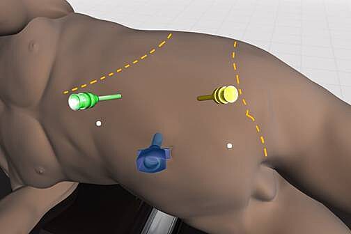
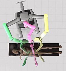
During laparoscopic partial nephrectomy approximately 3 to 5 small keyhole (< 1cm) incisions are made in the abdomen (Figure 2), which allow the surgeon to insert a telescope (called laparoscope) and hand held surgical instruments into the abdomen through portals call trocars.
The laparoscope allows for 10X magnification of the operative field, allowing the surgeon to accomplish the surgical procedure with improved visualization and without placing his hands into the abdominal cavity. The abdomen is filled with carbon dioxide gas to create a larger working space for the surgeon to accomplish the operation. This gas is later evacuated from the abdomen at the conclusion of the operation.
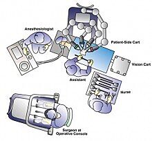
With the robotic technique, the da Vinci S Surgical Robotic System is assembled to the trocars prior to commencing the operation (Figure 3).
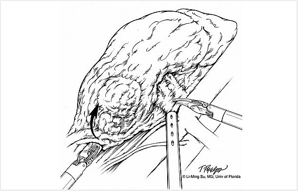
With the operating surgeon seated a few feet away at the surgeon operating console, the robotic instrumentation is controlled by the surgeon in real time with highly precise motion scaling (Figure 4).
The surgeon controls 2-3 multi-jointed robotic instruments to accomplish the tasks of dissection, cauterization, cutting and suturing (Figure 5).
In addition, the surgeon controls a stereoscopic lens which provides a three dimensional, high definition image of the anatomy. The affected kidney is then dissected and exposed. The tumor is then located and visualized in real time within the kidney with the use of a laparoscopic ultrasound probe. This ultrasound allows for precise delineation of the borders of the tumor within the kidney and for planning the proper line of excision in order to remove the mass completely. The blood supply to the kidney is temporarily clamped to minimize blood loss during excision of the tumor. The tumor, surrounding fat and a rim of healthy kidney is excised along with any visible surrounding lymph nodes. Once the tumor is excised from the kidney, it is immediately placed within a plastic sack which is later removed intact at the end of the operation through an extension of one of the existing abdominal incision sites. The kidney defect is then closed with sutures and surgical glue and the blood supply to the kidney restored. A small drain is left at the end of the procedure exiting one of the keyhole incisions. Finally the mass is removed from the abdomen through the plastic entrapment sack and the skin incisions closed using plastic surgery techniques to minimize scarring.
Slideshow
The following is a slide show of a series of schematic drawings to help patients better understand the steps involved with nerve-sparing robotic prostatectomy.
Full Procedural Video
Click to Individual Steps Below
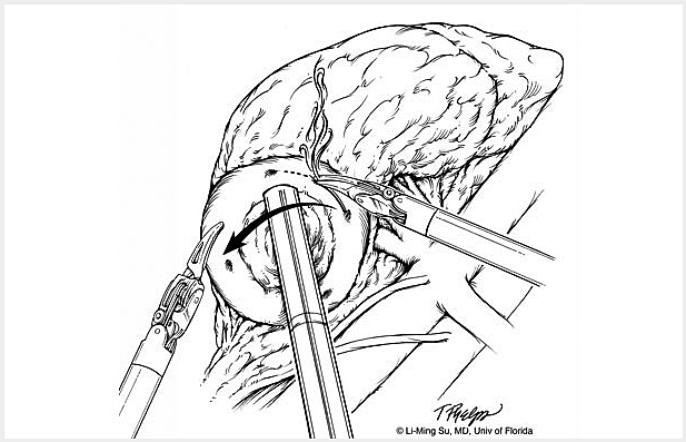
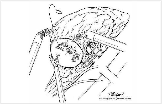
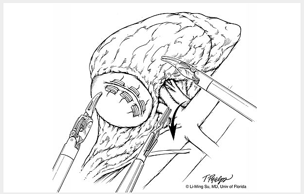
- Removal of Fat Overlying Renal Mass
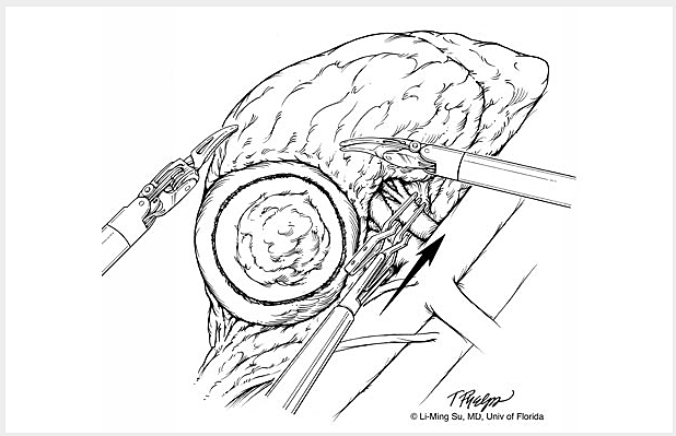
- Ultrasound of Renal Mass
- Demarcation of Renal Mass Margin
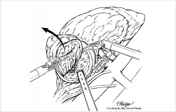
- Excision of Renal Mass
- Entrapment of Specimen
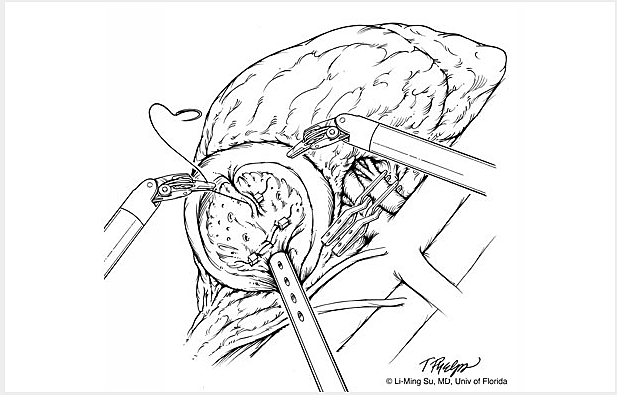
- Cauterization and Sutured Closure of Renal Defect
- Remove Clamp from Artery
- Place Drain
Potential Risks and Complications
As with any major surgery, complications, although rare, may occur with laparoscopic and robotic partial nephrectomy. Potential risks and complications with this operation include but are not limited to the following:
- Bleeding: Blood loss during this procedure is typically less than 100 cc with the rare need for a blood transfusion (<2% of patients). If you are interested in autologous blood transfusion (donating your own blood) prior to your surgery, you must make your surgeon aware. This can be arranged locally in Gainesville, FL at the Civitan Regional Blood center or at your local Red Cross.
- Infection: Although patients are given broad spectrum intravenous antibiotics immediately prior to surgery, infections of the urinary tract and skin incisions may still occur but are rare. If you develop any signs or symptoms of infection after the surgery (fever, drainage from or redness around your incisions, urinary frequency/discomfort, pain) please contact us at once.
- Urine Leak: When performing partial nephrectomy for large or deep seated tumors, the collecting system that drains the kidney of urine may be entered during surgical excision of the mass. Although the defect in the collecting system is closed with sutures, small leakage of urine from this site into the surrounding area of the kidney may occur. In the rare event of a large urine leak, a ureteral stent and/or urethral catheter may be required to allow for spontaneous resolution of the leak.
- Adjacent Tissue / Organ Injury: Although uncommon, adjacent organs and tissues may be injured as a result of your surgery. This includes the colon, bowel, vascular structures, nerves, muscles, spleen, liver, pancreas and gallbladder. If injury to your lung cavity occurs, a small chest tube may be required to evacuate air, blood, and fluid from around your lung, thus allowing your lung to expand and work properly. On rare occasions, further surgery may be required to address unexpected injuries to adjacent organs.
- Complete Removal of Kidney: In very rare cases, circumstances may arise that may lead your surgeon to remove the entire kidney. These include excessive bleeding, or a tumor that appears larger or more invasive than was appreciated on preoperative imaging tests. In such cases, the kidney can generally be removed safely by laparoscopy and often does not require conversion to open surgery.
- Incisional Hernia: Because of the small laparoscopic incisions, hernias at these sites occur rarely. In addition, larger incisions are closed carefully prior to the completion of your surgery to minimize the risk of hernias.
- Conversion to Open Surgery: In the rare event of complications or due to difficulty in dissecting by means of laparoscopy or robotic surgery, conversion to open surgery is sometimes required. This could result in a larger standard open incision and possibly a longer recuperation period.
What to Expect After Surgery
After a period of recovery in the Recovery Room, you will be transported to your hospital room once you are aware and your vital signs stable.
- Postoperative Pain: Although most patients in the first few days after surgery experience mild pain at their incision sites, this is generally well controlled by use of intravenous pain medication, patient-controlled anesthesia pump, or oral pain medication provided by your nurse. You may experience some minor transient shoulder pain (1-2 days) related to the carbon dioxide gas used to inflate your abdomen during the laparoscopic or robotic surgery.
- Nausea: Nausea can occur following any surgery especially those procedures that require general anesthesia. This is usually transient and controlled by medication which can be administered on an as needed basis by your nurse.
- Urinary Catheter: A urinary catheter (also called foley catheter) is placed to drain your bladder at the time of surgery while you are asleep. This is in efforts to monitor your urine output over the first day or so following surgery. This is generally removed by your nurse once you are walking comfortably on the first postoperative day. It is not uncommon to have blood-tinged urine for a few days after your surgery while your catheter is in place.
- Flank Drain: A small clear tube or drain will be placed during surgery exiting out of the side of your flank. The drain output will appear blood tinged but should be minimal. The drain primarily serves to identify any excessive bleeding or a urine leak from the partial nephrectomy site. The drain is typically removed on the day you are discharged from the hospital if the output remains low.
- Diet: Your diet will be advanced slowly following surgery from liquids to solids as tolerated. It is often the case that your appetite will be poor for up to a week following surgery. In addition, your intestinal function is often sluggish due to the effects of surgery and general anesthesia. It is for these two reasons that we recommend taking only small amounts of liquids by mouth at any one time until you begin to pass flatus and your appetite returns. In the meantime, your intravenous catheter will provide the necessary hydration to your body as you oral intake improves.
- Fatigue: Fatigue is quite common following surgery and should subside in a few weeks following surgery.
- Incentive Spirometry: You will be expected to do some very simple breathing exercises to help prevent respiratory infections through using an incentive spirometry device (these exercises will be explained to you by the nursing staff during your hospital stay). Coughing and deep breathing is an important part of your recuperation and helps prevent pneumonia and other pulmonary complications.
- Ambulation: On the evening of surgery it is very important to get out of bed and begin walking with the supervision of your nurse or family member to help prevent blood clots from forming in your legs. You can also expect to have SCD’s (sequential compression devices) wrapped around your lower legs and calf area to prevent blood clots called deep vein thrombosis from forming in your legs. In the days that follow surgery, patients are advised to walk at least 6 separate times a day in the hallways. This serves to further reduce the change of deep vein thrombosis and speed the return of bowel function.
- Constipation/Gas Cramps: You may experience sluggish bowels for several days following surgery as a result of the anesthesia. Suppositories and stool softeners are usually given to help with this problem. Taking a teaspoon of mineral oil daily at home will also help to prevent constipation. Narcotic pain medication can also cause constipation and therefore patients are encouraged to discontinue any narcotic pain medication as soon after surgery as tolerated.
- Hospital Stay: The length of hospital stay following laparoscopic and robotic partial nephrectomy is generally 1-2 days.
What to Expect After Discharge from the Hospital
- Pain Control: For the majority of patients, one to two days of oral narcotic pain medication may be necessary after which Extra Strength Tylenol is usually sufficient to manage their pain. Again, narcotics should be minimized to avoid constipation and over sedation.
- Showering: Patients can shower immediately upon discharge from the hospital allowing their incisions to get wet. Once out of the shower, pad your incision sites dry and avoid any heavy creams or lotions. Tub baths or hot tubs in the first 2 weeks are discouraged as this will allow for prolonged soaking of your incisions and increase the risk of infection. You may shower after returning home from the hospital. Your wound sites can get wet, but must be patted dry immediately after showering. The sutures underneath the skin will dissolve in 4-6 weeks.
- Activity: Walking 6 times a day for the first two weeks after surgery on a level surface is strongly encouraged as prolong sitting or lying can increase your risk of pneumonia and deep vein thrombosis. It is permissible to climb stairs. No heavy lifting or exertion for up to 4 weeks following surgery. Patients may begin driving once they are off of narcotic pain medication and have full range of motion at their waist. Most patients can return to full activity including work on an average 3-4 weeks after surgery.
- Diet: Patients may resume a regular diet once they begin to pass flatus and their appetite improves.
- Follow-up Appointment: Patients are routinely scheduled for their first postoperative appointment in the urology clinic approximately one month following surgery. To confirm your appointment time and date you can call the UF Health Urology – Medical Plaza at (352) 265-8240.
- Pathology Results: The pathology results from your surgery are usually available in approximately one week following surgery. Contact UF Health Urology – Medical Plaza at 352.265.8240 to obtain your results by phone. You are also welcome to request a copy of your pathology report through the Medical Records Department at (352) 265-0131.
Frequently Asked Questions (FAQ)
What is the advantage of laparoscopic and robotic partial nephrectomy as compared to open surgery?
These minimally invasive laparoscopic techniques have been routinely performed since 1992 and have translated into a significant benefit to patients including reduced blood loss and transfusions, reduced pain, shorter hospital stays, improved cosmesis, and a faster recovery as compared to open surgery. While open surgery requires either a large abdominal or flank incision, minimally invasive approaches involve 3-5 keyhole incisions in the abdomen. Published outcomes of laparoscopic and robotic partial nephrectomy demonstrate comparable cure rates to open partial nephrectomy.
Are there potential disadvantages?
Most patients with kidney tumors who are candidates for open surgery are also excellent candidates for a laparoscopic or robotic approach. These minimally invasive approaches have become the standard of care for most kidney tumors. In general there are no particular disadvantages; however, some situations may dictate the need for open surgery (see below).
What patients are not good candidates for laparoscopic and robotic partial nephrectomy?
Patients with very large tumors or tumors invading surrounding structures e.g. vena cava, liver, bowel may be best served by an open approach due to the extent and need for adjacent organ resection. Medical conditions such as severe lung and heart disease may not be able to tolerate a laparoscopic or robotic approach.
What is the difference between a laparoscopic and robotic approach?
Both are laparoscopic approaches and the choice of approach is a matter of surgeon preference. Operative times, blood loss, and hospital stays are similar between a pure laparoscopic and robotic technique. These procedures are performed by inflating the abdomen with carbon dioxide gas and placing a laparoscopic lens affixed to a high definition camera into the abdomen to view the internal organs. Conventional laparoscopic surgery involves hand held instruments, while robotic surgery involves the use of a sophisticated robotic device (called the da Vinci S Surgical Robotic System) with wristed instrumentation to allow the surgeon to dissect within the abdomen while controlling these instruments externally from a surgeon console.
What happens if complications arise and conversion to open surgery is required?
Although extremely rare, conversion to open surgery may be required if difficulty with dissection is encountered during the laparoscopic approach. Our surgeons are trained in open surgical approaches as well as laparoscopy and therefore are well equipped to complete the surgery in an open fashion if needed.
What is the overall success rate of laparoscopic and robotic partial nephrectomy?
Success rate in complete removal of the kidney tumor is similar to open surgical approaches. Prognosis of cancer-free survival is based upon the grade, stage and particular type of your cancer and will be discussed with you by your surgeon following surgery during review of the pathology report.
Will I need further treatment such as radiation or chemotherapy following surgery?
For patients with small, incidentally detected tumors on CT or MRI, prognosis remains excellent as most are cured with surgery alone. Rarely, patients are found to have large, invasive cancers that may require adjuvant treatment with medical therapies such as interleukin-2, interferon-alpha, or tyrosine kinase inhibitors. These would be administrated under advisement of a medical oncologist. Currently there is no utility for radiation or chemotherapy.