Lumbosacral spine x-ray
Definition
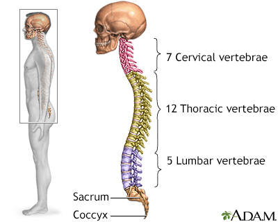
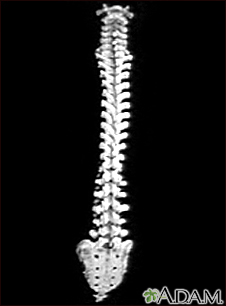
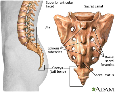
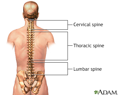
A lumbosacral spine x-ray is a picture of the small bones (vertebrae) in the lower part of the spine. This area includes the lumbar region and the sacrum, the area that connects the spine to the pelvis.
Alternative Names
X-ray - lumbosacral spine; X-ray - lower spine
How the Test is Performed
The test is done in a hospital x-ray department or your health care provider's office by an x-ray technician. You will be asked to lie on the x-ray table in different positions. If the x-ray is being done to diagnose an injury, care will be taken to prevent further injury.
The x-ray machine will be placed over the lower part of your spine. You will be asked to hold your breath as the picture is taken so that the image will not be blurry. In most cases, 3 to 5 pictures are taken.
How to Prepare for the Test
Tell the provider if you are pregnant. Take off all jewelry.
How the Test will Feel
There is rarely any discomfort when having an x-ray, although the table may be cold.
Why the Test is Performed
Often, the provider will treat a person with low back pain for 4 to 8 weeks before ordering an x-ray.
The most common reason for lumbosacral spine x-ray is to look for the cause of low back pain that:
- Occurs after injury
- Is severe
- Does not go away after 4 to 8 weeks
- Is present in an older person
What Abnormal Results Mean
Lumbosacral spine x-rays may show:
- Abnormal curves of the spine
- Abnormal wear on the cartilage and bones of the lower spine, such as bone spurs and narrowing of the joints between the vertebrae
- Cancer (although cancer often cannot be seen on this type of x-ray)
- Fractures
- Signs of thinning bones (osteoporosis)
- Spondylolisthesis, in which a bone (vertebra) in the lower part of the spine slips out of the proper position onto the bone below it
Though some of these findings may be seen on an x-ray, they are not always the cause of back pain.
Many problems in the spine cannot be diagnosed using a lumbosacral x-ray, including:
- Sciatica
- Slipped or herniated disc
- Spinal stenosis - narrowing of the spinal column
Risks
There is low radiation exposure. X-ray machines are checked often to make sure they are as safe as possible. Most experts feel that the risk is low compared with the benefits.
Pregnant women should not be exposed to radiation, if at all possible. Care should be taken before children receive x-rays.
Considerations
There are some back problems that an x-ray will not find. That is because they involve the muscles, nerves, and other soft tissues. A lumbosacral spine CT or lumbosacral spine MRI are better options for soft tissue problems.
Gallery
References
Contreras F, Perez J, Jose J. Imaging overview. In: Miller MD, Thompson SR, eds. DeLee, Drez, & Miller's Orthopaedic Sports Medicine. 5th ed. Philadelphia, PA: Elsevier; 2020:chap 7.
Kapoor G, Toms AP. Current status of imaging of the musculoskeletal system. In: Adam A, Dixon AK, Gillard JH, Schaefer-Prokop CM, eds. Grainger & Allison's Diagnostic Radiology: A Textbook of Medical Imaging. 7th ed. Philadelphia, PA: Elsevier; 2021:chap 38.
Parizel PM, Van Thielen T, van den Hauwe L, Van Goethem JW. Degenerative disease of the spine. In: Adam A, Dixon AK, Gillard JH, Schaefer-Prokop CM, eds. Grainger & Allison's Diagnostic Radiology: A Textbook of Medical Imaging. 7th ed. Philadelphia, PA: Elsevier; 2021:chap 48.
Warner WC, Sawyer JR. Scoliosis and kyphosis. In: Azar FM, Beaty JH, eds. Campbell's Operative Orthopaedics. 14th ed. Philadelphia, PA: Elsevier; 2021:chap 44.