Mitral valve surgery - minimally invasive
Definition
Mitral valve surgery is surgery to either repair or replace the mitral valve in your heart.
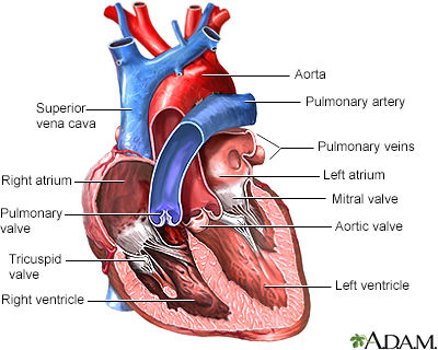
Blood flows from the lungs and enters a pumping chamber of the heart called the left atrium. The blood then flows into the final pumping chamber of the heart called the left ventricle. The mitral valve is located between these two chambers. It makes sure that the blood keeps moving forward through the heart.
You may need surgery on your mitral valve if:
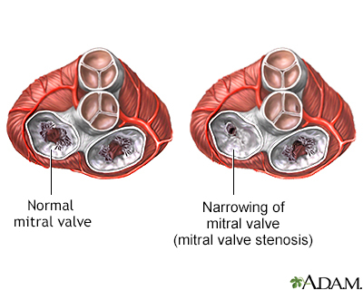
- The mitral valve is hardened (calcified). This prevents blood from moving forward through the valve.
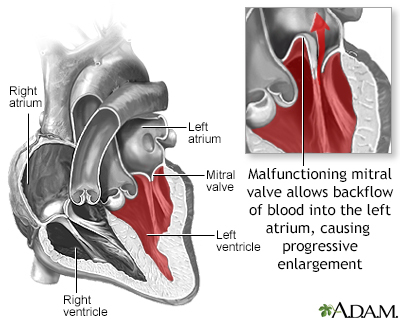
- The mitral valve is too loose. Blood tends to flow backward when this occurs.
Minimally invasive mitral valve surgery is done through several small cuts. Another type of operation, open mitral valve surgery, requires a larger cut.
Alternative Names
Mitral valve repair - right mini-thoracotomy; Mitral valve repair - partial upper or lower sternotomy; Robotically-assisted endoscopic valve repair; Percutaneous mitral valvuloplasty
Description
Before your surgery, you will receive general anesthesia.
You will be asleep and pain-free.
There are several different ways to perform minimally invasive mitral valve surgery.
- Your heart surgeon may make a 2-inch to 3-inch-long (5 to 7.5 centimeters) cut in the right part of your chest near the sternum (breastbone). Muscles in the area will be divided. This lets the surgeon reach the heart. A small cut is made in the left side of your heart so the surgeon can repair or replace the mitral valve.
- In endoscopic surgery, your surgeon makes 1 to 4 small holes in your chest. Surgery is done through the cuts using a camera and special surgical tools. For robotically-assisted valve surgery, the surgeon makes 2 to 4 tiny cuts in your chest. The cuts are about 1/2 to 3/4 inches (1.5 to 2 centimeters) each. The surgeon uses a special computer to control robotic arms during the surgery. A 3D view of the heart and mitral valve are displayed on a computer in the operating room.
You will need a heart-lung machine for these types of surgery. You will be connected to this device through small cuts in the groin or on the chest.
If your surgeon can repair your mitral valve, you may have:
- Ring annuloplasty -- The surgeon tightens the valve by sewing a ring of metal, cloth, or tissue around the valve.
- Valve repair -- The surgeon trims, shapes, or rebuilds one or both of the flaps that open and close the valve.
You will need a new valve if there is too much damage to your mitral valve. This is called replacement surgery. Your surgeon may remove some or all of your mitral valve and sew a new one into place. There are two main types of new valves:
- Mechanical -- Made of man-made materials, such as titanium and carbon. These valves last the longest. You will need to take blood-thinning medicine, such as warfarin (Coumadin), for the rest of your life.
- Biological -- Made of human or animal tissue. These valves last 10 to 15 years or longer, but you will probably not need to take blood thinners for life.
The surgery may take 2 to 4 hours.
This surgery can sometimes be done through a groin artery, with no cuts on your chest. The doctor sends a catheter (flexible tube) with a balloon attached on the end. The balloon inflates to stretch the opening of the valve. This procedure is called percutaneous valvuloplasty and done for a blocked mitral valve.
A new procedure involves placing a catheter through an artery in the groin and clipping the valve to prevent the valve from leaking.
Why the Procedure Is Performed
You may need surgery if your mitral valve does not work properly because:
- You have mitral regurgitation -- When a mitral valve does not close all the way and allows blood to leak back into the left atria.
- You have mitral stenosis -- When a mitral valve does not open fully and restricts blood flow.
- Your valve has developed an infection (infectious endocarditis).
- You have severe mitral valve prolapse that is not controlled with medicine.
Minimally invasive surgery may be done for these reasons:
- Changes in your mitral valve are causing major heart symptoms, such as shortness of breath, leg swelling, or heart failure.
- Tests show that the changes in your mitral valve are beginning to harm your heart function.
- Damage to your heart valve from infection (endocarditis).
A minimally invasive procedure has many benefits. There is less pain, blood loss, and risk of infection. You will also recover faster than you would from open heart surgery. However, some people may not be able to have this type of procedure.
Percutaneous valvuloplasty can only be done in people who are too sick to have anesthesia. The results of this procedure are not long-lasting.
Risks
Risks for any surgery are:
- Blood clots in the legs that may travel to the lungs
- Blood loss
- Breathing problems
- Infection, including in the lungs, kidneys, bladder, chest, or heart valves
- Reactions to medicines
Minimally invasive surgery techniques have far fewer risks than open surgery. Possible risks from minimally invasive valve surgery are:
- Damage to other organs, nerves, or bones
- Heart attack, stroke, or death
- Infection of the new valve
- Irregular heartbeat that must be treated with medicines or a pacemaker
- Kidney failure
- Poor healing of the wounds
Before the Procedure
Always tell your health care provider:
- If you are or could be pregnant
- What medicines you are taking, even drugs, supplements, or herbs you bought without a prescription
You may be able to store blood in the blood bank for transfusions during and after your surgery. Ask your provider about how you and your family members can donate blood.
If you smoke, you should stop. Ask your provider for help.
During the days before your surgery:
- For the 1 week period before surgery, you may be asked to stop taking medicines that make it harder for your blood to clot. These might cause increased bleeding during the surgery. Some of these medicines include aspirin, ibuprofen (Advil, Motrin), and naproxen (Aleve, Naprosyn).
- If you are taking warfarin (Coumadin) or clopidogrel (Plavix), talk with your surgeon before stopping or changing how you take these drugs.
- Ask which drugs you should still take on the day of your surgery.
- Prepare your house for when you get home from the hospital.
- Shower and wash your hair the day before surgery. You may need to wash your body below your neck with a special soap. Scrub your chest 2 or 3 times with this soap. You also may be asked to take an antibiotic to prevent infection.
On the day of the surgery:
- You may be asked not to drink or eat anything after midnight the night before your surgery. This includes using chewing gum and mints. Rinse your mouth with water if it feels dry. Be careful not to swallow.
- Take the medicines you have been told to take with a small sip of water.
- You will be told when to arrive at the hospital.
After the Procedure
Expect to spend 3 to 5 days in the hospital after surgery. You will wake up in the intensive care unit (ICU) and recover there for 1 or 2 days. Nurses will closely watch monitors that display your vital signs (pulse, temperature, and breathing).
Two to three tubes will be in your chest to drain fluid from around your heart. They are usually removed 1 to 3 days after surgery. You may have a catheter (flexible tube) in your bladder to drain urine. You may also have intravenous (IV) lines to get fluids.
You will go from the ICU to a regular hospital room. Your heart and vital signs will be monitored until you are ready to go home. You will receive pain medicine for pain in your chest.
Your nurse will help start activity slowly. You may begin a program to make your heart and body stronger.
A pacemaker may be placed in your heart if your heart rate becomes too slow after surgery. This may be temporary or you may need a permanent pacemaker before you leave the hospital.
Outlook (Prognosis)
Mechanical heart valves do not fail often. However, blood clots can develop on them. If a blood clot forms, you may have a stroke. Bleeding can occur, but this is rare.
Biological valves have a lower risk of blood clots, but tend to fail over a long period of time.
The results of mitral valve repair are excellent. For best results, choose to have surgery at a center that does many of these procedures. Minimally invasive heart valve surgery has improved greatly in recent years. These techniques are safe for most people, and can reduce recovery time and pain.
Gallery
References
Bajwa G, Mihaljevic T. Minimally invasive mitral valve surgery: partial sternotomy approach. In: Sellke FW, Ruel M, eds. Atlas of Cardiac Surgical Techniques. 2nd ed. Philadelphia, PA: Elsevier; 2019:chap 20.
Goldstone AB, Woo YJ. Surgical treatment of the mitral valve. In: Sellke FW, del Nido PJ, Swanson SJ, eds. Sabiston and Spencer Surgery of the Chest. 9th ed. St Louis, MO: Elsevier; 2016:chap 80.
Herrmann HC, Mack MJ. Transcatheter therapies for valvular heart disease. In: Zipes DP, Libby P, Bonow RO, Mann DL, Tomaselli GF, Braunwald E, eds. Braunwald's Heart Disease: A Textbook of Cardiovascular Medicine. 11th ed. Philadelphia, PA: Elsevier; 2019:chap 72.
Thomas JD, Bonow RO. Mitral valve disease. In: Zipes DP, Libby P, Bonow RO, Mann DL, Tomaselli GF, Braunwald E, eds. Braunwald's Heart Disease: A Textbook of Cardiovascular Medicine. 11th ed. Philadelphia, PA: Elsevier; 2019:chap 69.
Related specialties
Aftercare and more
Our experts

Community and Patient Programs: Mitral valve surgery - minimally invasive
Our community and patient programs provide great value to patients, families and loved ones. People can find support, educational materials, expert consultants and more. In most instances, these programs are offered free of charge.
-
Preparing Your Child for Hospital Procedures
Designed to help you and your child prepare for common hospital procedures.