Pupil - white spots
Definition
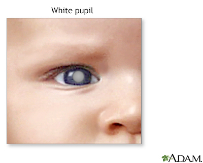
White spots in the pupil is a condition that causes the pupil of the eye to look white instead of black.
Alternative Names
Leukocoria
Considerations
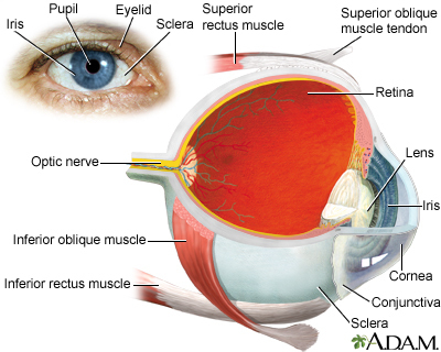
The pupil of the human eye is normally black. In flash photographs the pupil may appear red. This is called the "red reflex" by health care providers and is normal.
Sometimes, the pupil of the eye may appear white, or the normal red reflex may appear to be white. This is not a normal condition, and you need to see an eye care provider right away.
There are many different causes of white pupil or white reflex. Other conditions also can mimic white pupil. If the cornea, which is normally clear, becomes cloudy, it may look similar to a white pupil. Although the causes of a cloudy or white cornea are different from those of a white pupil or white reflex, these problems also need medical attention right away.
Cataracts may also cause the pupil to appear white.
Causes
Causes of this condition may include:
- Coats disease - exudative retinopathy
- Coloboma
- Congenital cataract (may be hereditary or may result from other conditions, including congenital rubella, galactosemia, retrolental fibroplasia)
- Persistent primary hyperplastic vitreous
- Retinoblastoma
- Toxocara canis (infection caused by a parasite)
- Uveitis
Home Care
Most causes of white pupil will cause decreased vision. This may often occur before the pupil appears to be white.
Detecting a white pupil is especially important in infants. Babies are unable to communicate to others that their vision is decreased. It is also harder to measure an infant's vision during an eye exam.
If you see a white pupil, call your provider right away. Well-child exams routinely screen for a white pupil in children. A child that develops a white pupil or cloudy cornea needs immediate attention, preferably from an eye specialist.
It is important to get diagnosed early if the problem is caused by retinoblastoma since this disease can be fatal.
When to Contact a Medical Professional
Contact your provider if you notice any color changes in the pupil or cornea of the eye.
What to Expect at Your Office Visit
The provider will do a physical exam and ask about your symptoms and medical history.
The physical exam will include a detailed eye examination.
The following tests may be performed:
Gallery
References
Cioffi GA, LIebmann JM. Diseases of the visual system. In: Goldman L, Schafer AI, eds. Goldman-Cecil Medicine. 26th ed. Philadelphia, PA: Elsevier; 2020:chap 395.
Olitsky SE, Marsh JD. Abnormalities of the pupil and iris. In: Kliegman RM, St. Geme JW, Blum NJ, Shah SS, Tasker RC, Wilson KM, eds. Nelson Textbook of Pediatrics. 21st ed. Philadelphia, PA: Elsevier; 2020:chap 640.
Vagge A, Wangtiraumnuay N, Pellegrini M, Scotto R, Iester M, Traverso CE. Evaluation of a free public smartphone application to detect leukocoria in high-risk children aged 1 to 6 years. J Pediatr Ophthalmol Strabismus. 2019;56(4):229-232.PMID: 31322712 pubmed.ncbi.nlm.nih.gov/31322712/.