Solitary pulmonary nodule
Definition
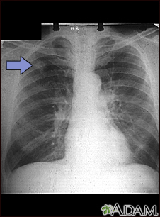
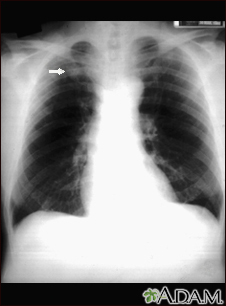
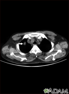
A solitary pulmonary nodule is a round or oval spot (lesion) in the lung that is seen with a chest x-ray or CT scan.
Alternative Names
Lung cancer - solitary nodule; Infectious granuloma - pulmonary nodule; SPN
Causes
More than half of all solitary pulmonary nodules are noncancerous (benign). Benign nodules have many causes, including scars and past infections.
Infectious granulomas (which are formed by cells as a reaction to a past infection) cause most benign lesions. Common infections that often result in granulomas or other healed scars include:
- Tuberculosis (TB) or exposure to TB
Primary lung cancer is the most common cause of cancerous (malignant) pulmonary nodules. This is cancer that starts in the lung.
Symptoms
A solitary pulmonary nodule itself rarely causes symptoms.
Exams and Tests
A solitary pulmonary nodule is most often found on a chest x-ray or chest CT scan. These imaging tests are often done for other symptoms or reasons.
Your health care provider must decide whether the nodule in your lung is most likely benign or of concern. A nodule more is likely benign if:
- The nodule is small, has a smooth border, and has a solid and even appearance on an x-ray or CT scan.
- You are young and do not smoke.
Your provider may then choose to monitor the nodule over time by repeating a series of x-rays or CT scans.
- Repeat chest x-rays or chest CT scans are the most common way to monitor the nodule. Sometimes, lung PET scans may be done.
- If repeated x-rays show that the nodule size has not changed in 2 years, it is most likely benign and a biopsy is not needed.
Your provider may choose to biopsy the nodule to rule out cancer if:
- You are a smoker.
- You have other symptoms of lung cancer.
- The nodule has grown in size or has changed when compared to earlier images.
A lung needle biopsy may be done by placing a needle directly through the wall of your chest, or during procedures called bronchoscopy or mediastinoscopy.
Tests to rule out TB and other infections may also be done.
Treatment
Ask your provider about the risks of having a biopsy versus monitoring the size of the nodule with regular x-rays or CT scans. Treatment may be based on the results of the biopsy or other tests.
Outlook (Prognosis)
The outlook is usually good if the nodule is benign. If the nodule does not grow larger over a 2-year period, often nothing more needs to be done.
Gallery
References
Bueno J, Landeras L, Chung JH. Updated Fleischner Society guidelines for managing incidental pulmonary nodules: common questions and challenging scenarios. Radiographics. 2018;38(5):1337-1350. PMID: 30207935 pubmed.ncbi.nlm.nih.gov/30207935/.
Jokerst CE, Gotway MB. Thoracic radiology: noninvasive diagnostic imaging. In: Broaddus VC, Ernst JD, King TE, et al, eds. Murray and Nadel's Textbook of Respiratory Medicine. 7th ed. Philadelphia, PA: Elsevier; 2022:chap 20.
Reed JC. Solitary pulmonary nodule. In: Reed JC, ed. Chest Radiology: Patterns and Differential Diagnoses. 7th ed. Philadelphia, PA: Elsevier; 2018:chap 20.
