Standard eye exam
Definition
A standard eye exam is a series of tests done to check your vision and the health of your eyes.
Alternative Names
Standard ophthalmic exam; Routine eye examination; Eye exam - standard; Annual eye exam
How the Test is Performed
First, you will be asked if you are having any eye or vision problems. You will be asked to describe these problems, how long you have had them, and any factors that have made them better or worse.
Your history of glasses or contact lenses will also be reviewed. The eye doctor will then ask about your overall health, including any medicines you take and your family's medical history.
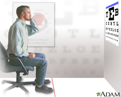
Next, the doctor will check your vision (visual acuity) using a Snellen chart.
- You will be asked to read random letters that become smaller line by line as your eyes move down the chart. Some Snellen charts are actually video monitors showing letters or images.
- To see if you need glasses, the doctor will place several lenses in front of your eye, one at a time, and ask you when the letters on the Snellen chart become easier to see. This is called a refraction.
Other parts of the exam include tests to:
- See if you have proper three-dimensional (3D) vision (stereopsis).
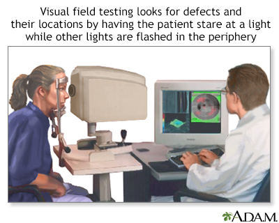
- Check your side (peripheral) vision.
- Check the eye muscles by asking you to look in different directions at a penlight or other small object.
- Examine the pupils with a penlight to see if they respond (constrict) properly to light.
- Often, you'll be given eye drops to open up (dilate) your pupils. This allows the doctor to use a device called an ophthalmoscope to view the structures at the back of the eye. This area is called the fundus. It includes the retina and nearby blood vessels and optic nerve.
Another magnifying device, called a slit lamp, is used to:
- See the front parts of the eye (eyelids, cornea, conjunctiva, sclera, and iris)
- Check for increased pressure in the eye (glaucoma) using a method called tonometry
Color blindness is tested using cards with colored dots that form numbers.
How to Prepare for the Test
Make an appointment with an eye doctor (some take walk-in patients). Avoid eye strain on the day of the test. If you wear glasses or contacts, bring them with you. You may need someone to drive you home if the doctor uses eye drops to dilate your pupils.
How the Test will Feel
The tests cause no pain or discomfort.
Why the Test is Performed
All children should have vision screening in a pediatrician's or family practitioner's office around the time when they learn the alphabet, and then every 1 to 2 years afterward. Screening should begin sooner if any eye problems are suspected.
Between ages 20 and 39:
- A complete eye exam should be done every 5 to 10 years
- Adults who wear contact lenses need yearly eye exams
- Certain eye symptoms or disorders may require more frequent exams
Adults over age 40 who have no risk factors or ongoing eye conditions should be screened:
- Every 2 to 4 years for adults ages 40 to 54
- Every 1 to 3 years for adults ages 55 to 64
- Every 1 to 2 years for adults age 65 and older
Depending on your risk factors for eye diseases and your current symptoms or illnesses, your eye doctor may recommend that you have exams more often.
Some of the eye and medical problems that can be found by a routine eye test include:
- Cataracts (clouding of the lens of the eye)
- Diabetes
- Glaucoma
- High blood pressure
- Age-related macular degeneration (ARMD), (loss of sharp, central vision)
Normal Results
Results of a routine eye exam are normal when the eye doctor finds you have:
- 20/20 (6/6) (normal) vision
- Ability to identify different colors
- Full visual field
- Proper eye muscle coordination
- Normal eye pressure
- Normal eye structures (cornea, iris, lens)
What Abnormal Results Mean
Abnormal results may be due to any of the following:
- ARMD
- Astigmatism (abnormally curved cornea)
- Blocked tear duct
- Cataracts
- Color blindness
- Corneal dystrophy
- Corneal ulcers, infections, or injury
- Damaged nerves or blood vessels in the eye
- Diabetes-related damage in the eye (diabetic retinopathy)
- Hyperopia (farsightedness)
- Glaucoma
- Injury of the eye
- Lazy eye (amblyopia)
- Myopia (nearsightedness)
- Presbyopia (inability to focus on near objects that develops with age)
- Strabismus (crossed eyes)
- Retinal tear or detachment
This list may not include all possible causes of abnormal results.
Risks
If you receive drops to dilate your eyes for the ophthalmoscopy, your vision will be blurred.
- Wear sunglasses to protect your eyes from sunlight, which can damage your eyes more when they are dilated.
- Have someone drive you home.
- The drops usually wear off in several hours.
In rare cases, the dilating eyedrops cause:
- An attack of narrow-angle glaucoma
- Dizziness
- Dryness of the mouth
- Flushing
- Nausea and vomiting
Gallery
References
Ball JW, Dains JE, Flynn JA, Solomon BS, Stewart RW. Eyes. In: Ball JW, Dains JE, Flynn JA, Solomon BS, Stewart RW, eds. Seidel's Guide to Physical Examination. 9th ed. St Louis, MO: Elsevier; 2019:chap 12.
Chuck RS, Dunn SP, Flaxel CJ; American Academy of Ophthalmology Preferred Practice Pattern Committee, et al. Comprehensive adult medical eye evaluation preferred practice pattern. Ophthalmology. 2021;128(1):1-29. www.aaojournal.org/article/S0161-6420(20)31026-5/fulltext. Published November 12, 2020. Accessed March 2, 2021.
Olitsky SE, Marsh JD. Examination of the eye. In: Kliegman RM, St. Geme JW, Blum NJ, Shah SS, Tasker RC, Wilson KM, eds. Nelson Textbook of Pediatrics. 21st ed. Philadelphia, PA: Elsevier; 2020:chap 637.
Prokopich CL, Hrynchak P, Flanagan JG, Hynes AF, Chisholm C. Ocular health assessment. In: Elliott DB, ed. Clinical Procedures in Primary Eye Care. 5th ed. Philadelphia, PA: Elsevier; 2021:chap 7.