Definition
Subconjunctival hemorrhage is a bright red patch appearing in the white of the eye. This condition is one of several disorders called red eye.
Causes
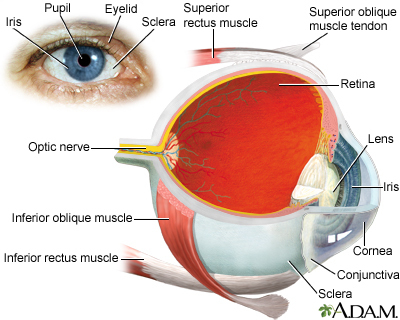
The white of the eye (sclera) is covered with a thin layer of clear tissue called the bulbar conjunctiva. A subconjunctival hemorrhage occurs when a small blood vessel breaks open and bleeds within the conjunctiva. The blood is often very visible, but since it is confined within the conjunctiva, it does not move and cannot be wiped away. The problem may occur without injury. It is often first noticed when you wake up and look in a mirror.
Some things that may cause a subconjunctival hemorrhage include:
- Sudden increases in pressure, such as violent sneezing or coughing
- Having high blood pressure or taking blood thinners
- Rubbing the eyes
- Viral infection
- Certain eye surgeries or injuries
A subconjunctival hemorrhage is common in newborn infants. In this case, the condition is thought to be caused by the pressure changes across the infant's body during childbirth.
Symptoms
A bright red patch appears on the white of the eye. The patch does not cause pain and there is no discharge from the eye. Vision does not change.
Exams and Tests
The health care provider will perform a physical exam and look at your eyes.
Blood pressure should be tested. If you have other areas of bleeding or bruising, more specific tests may be needed.
Treatment
No treatment is needed. You should have your blood pressure checked regularly.
Outlook (Prognosis)
A subconjunctival hemorrhage most often goes away on its own in about 2 to 3 weeks. The white of the eye may look yellow as the problem goes away.
Possible Complications
In most cases, there are no complications. Rarely, a total subconjunctival hemorrhage may be a sign of a serious vascular disorder in older people.
When to Contact a Medical Professional
Call your provider if a bright red patch appears on the white of the eye.
Prevention
There is no known prevention.
References
Dorsch JN. Red eye. In: Kellerman RD, Rakel DP, eds. Conn's Current Therapy 2021. Philadelphia, PA: Elsevier 2021:509-513.
Guluma K, Lee JE. Ophthalmology. In: Walls RM, Hockberger RS, Gausche-Hill M, eds. Rosen's Emergency Medicine: Concepts and Clinical Practice. 9th ed. Philadelphia, PA: Elsevier; 2018:chap 61.
Salmon JF. Conjunctiva. In: Salmon JF, ed. Kanski's Clinical Ophthalmology. 9th ed. Philadelphia, PA: Elsevier; 2020:chap 6.