Update Location

My Location

Update your location to show providers, locations, and services closest to you.

Enter a zip code
Or
Select a campus/region

Definition

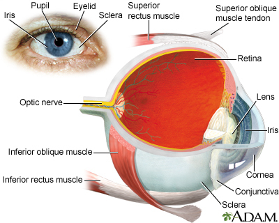
The uvea is the middle layer of the eye. It lies beneath the white part of the eye (the sclera). It is made of the iris, ciliary body, and choroid. These structures control many eye functions, including adjusting to different levels of light or distances of objects. Inflammation of one or more of these structures is called uveitis.

Alternative Names

Vascular tunic

Gallery

Eye
The eye is the organ of sight, a nearly spherical hollow globe filled with fluids (humors). The outer layer or tunic (sclera, or white, and cornea) is fibrous and protective. The middle layer (choroid, ciliary body and the iris) is vascular. The innermost layer (the retina) is nervous or sensory. The fluids in the eye are divided by the lens into the vitreous humor (behind the lens) and the aqueous humor (in front of the lens). The lens itself is flexible and suspended by ligaments which allow it to change shape to focus light on the retina, which is composed of sensory neurons.

References

Evans M. Anatomy of the uvea. In: Yanoff M, Duker JS, eds. Ophthalmology. 5th ed. Philadelphia, PA: Elsevier; 2019:chap 7.1.

Taber's Cyclopedic Medical Dictionary. 24th ed. F.A. Davis Company; 2021 www.tabers.com/tabersonline. Accessed April 5, 2022.

Last reviewed November 4, 2021 by Linda J. Vorvick, MD, Clinical Associate Professor, Department of Family Medicine, UW Medicine, School of Medicine, University of Washington, Seattle, WA. Also reviewed by David Zieve, MD, MHA, Medical Director, Brenda Conaway, Editorial Director, and the A.D.A.M. Editorial team..

Related specialties