Venogram - leg
Definition
Venography for legs is a test used to see the veins in the leg.
X-rays are a form of electromagnetic radiation, like visible light is. However, these rays are of higher energy. Therefore, they can go through the body to form an image on film. Structures that are dense (such as bone) will appear white, air will be black, and other structures will be shades of gray.
Veins are not normally seen in an x-ray, so a special dye is used to highlight them. This dye is called contrast.
Alternative Names
Phlebogram - leg; Venography - leg; Angiogram - leg
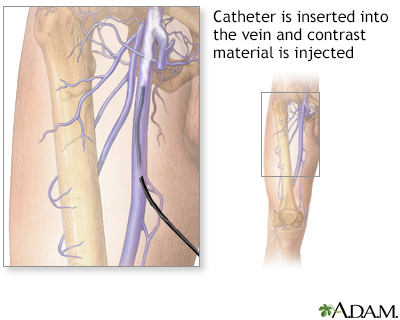
How the Test is Performed
This test is usually done in a hospital. You will be asked to lie on an x-ray table. A numbing medicine is applied to the area. You may ask for a sedative if you are anxious about the test.
The health care provider places a needle into a vein in the foot of the leg being looked at. An intravenous (IV) line is inserted through the needle. The contrast dye flows through this line into the vein. A tourniquet may be placed on your leg so the dye flows into the deeper veins.
X-rays are taken as the dye flows through the leg.
The catheter is then removed, and the puncture site is bandaged.
How to Prepare for the Test
You will wear hospital clothing during this procedure. You will be asked to sign a consent form for the procedure. Remove all jewelry from the area being imaged.
Tell the provider:
- If you are pregnant
- If you have allergies to any medicines
- Which medicines you are taking (including any herbal preparations)
- If you have ever had any allergic reactions to x-ray contrast material or iodine substance
How the Test will Feel
The x-ray table is hard and cold. You may want to ask for a blanket or pillow. You will feel a sharp poke when the intravenous catheter is inserted. As the dye is injected, you may experience a burning sensation.
There may be tenderness and bruising at the site of the injection after the test.
Why the Test is Performed
This test is used to identify and locate blood clots in the veins of the legs.
Normal Results
Free flow of the blood through the vein is normal.
What Abnormal Results Mean
Abnormal results may be due to a blockage. The blockage can be caused by:
- Blood clot
- Tumor
- Inflammation
Risks
Risks of this test are:
- Allergic reaction to the contrast dye
- Kidney failure, especially in the older adults or people with diabetes who take the medicine metformin (Glucophage)
- Worsening of a clot in the leg vein
There is low radiation exposure. However, most experts feel that the risk of most x-rays is smaller than other daily risks. Pregnant women and children are more sensitive to the risks of the x-ray.
Considerations
Ultrasound is used more often than this test because it has fewer risks and side effects. MRI and CT scans may also be used to look at the veins in the leg.
Gallery
References
Ameli-Renani S, Belli A-M, Chun J-Y, Morgan RA. Peripheral vascular disease intervention. In: Adam A, Dixon AK, Gillard JH, Schaefer-Prokop CM, eds. Grainger & Allison's Diagnostic Radiology: A Textbook of Medical Imaging. 7th ed. Philadelphia, PA: Elsevier; 2021:chap 80.
Bechara CF. Venography. In: Sidawy AN, Perler BA, eds. Rutherford's Vascular Surgery and Endovascular Therapy. 10th ed. Philadelphia, PA: Elsevier; 2023:chap 28.
