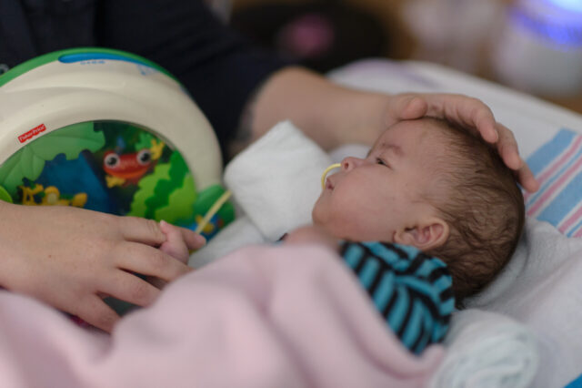
Conjoined twins connected at the heart and liver successfully separated at UF Health

[slideshow url="https://media.news.health.ufl.edu/slide/2016_09_21_Conjoined_Twins/index.html"]
GAINESVILLE, Fla. — Conjoined twin girls who were connected at the heart and other organs have been successfully separated in an extremely rare surgery performed by physicians at University Florida Health Shands Children’s Hospital.
The girls, who were born at UF Health Shands Hospital in April and separated in June, each had their own complete set of organs but were attached at the liver, diaphragm, sternum and heart, called a thoraco-omphalopagus connection. Their hearts were the most critical element of the separation, according to Mark Bleiweis, M.D., chief of pediatric and congenital cardiovascular surgery at UF Health and the surgeon who performed the heart separation. The twins shared a connection at the upper chamber of the heart, called the atrium, where blood enters the heart.
“It was a really complex connection because it was close to very important veins in the hearts of both babies,” Bleiweis said. “In the world, there have not been many successful separations with a cardiac connection. It became a very challenging planning process for us, and, ultimately, a challenging separation.”
Jennifer Co-Vu, M.D., FAAP, specializes in fetal cardiac care. She first studied the physiology of the unborn twins during an hourslong ultrasound in Jacquelyn’s 21st week of pregnancy and she told the parents she thought that not only would the babies survive birth, they also would survive after they were born. The parents had two options: to attempt separation, or to be prepared to raise conjoined twins, Co-Vu said.
Co-Vu and the team used cardiac CT and MRI scans before and after the twins were born to create what appears to be the first-ever 3-D printed conjoined twin heart. The replica allowed the surgeons to examine the shared structures in the heart and plan how to separate them. The team — which included Co-Vu, Bleiweis and many others from the pediatric cardiac intensive care unit, radiology, neonatology, maternal fetal medicine, anesthesiology and plastic surgery — met numerous times to create the preoperative plan for the twins, to practice the separation itself and to manage the twins’ care after the surgery.
“When I saw the heart structures and liver structures in utero, I had a feeling that we could separate them, but I had to examine the anatomy more closely and consult with my cardiology colleagues at the UF Health Congenital Heart Center,” said Co-Vu, director of the Fetal Cardiac Program. “I was able to give them hope, yet at the same time, I told them I was cautiously optimistic. … We are very fortunate that this was a success.”
Conjoined twins occur only in about 1 in 200,000 live births. Between 40 and 60 percent of conjoined twins are stillborn, and 35 percent who live through birth survive only one day, according to the University of Maryland Medical Center. Only about 5 to 25 percent of conjoined twins survive, and survival of twins connected at the heart is extremely rare. In cases where the hearts are joined, the decision is often made to not do separation surgery in cases where there is a connection at the heart.
In addition to being joined at the heart, the girls also shared a large, fused liver, according to Saleem Islam, M.D., M.P.H., chief of the division of pediatric surgery in the UF College of Medicine.
“The liver, from all of the imaging we obtained both before the babies’ birth and after they were born, indicated that it was almost like one giant liver without any true plane of separation,” Islam said.
Without a clear picture of how to separate the liver before the surgery took place, Islam and his team had to use a method called “intraoperative ultrasound” to guide the separation. Using this method, Islam looked for areas of the joined liver that were free from large blood vessels.
During the the procedure, which spanned about six to eight hours, a pediatric surgery team with two surgeons, a cardiothoracic surgery team with two surgeons, two pediatric cardiac anesthesiology teams, a pediatric cardiac imaging team, and multiple nursing and ancillary staff worked to safely and successfully separate the infants. To keep the various equipment keeping the twins alive during the surgery separated and easily identifiable, the staff wrapped tubing and electrical wiring with orange tape for one of the twins and blue tape for her sister, according to anesthesiologist Andrew Pitkin, MBBS, MRCP, FRCA.
Physicians at a different hospital discovered that mother Jacquelyn was carrying conjoined twins when the parents went for their first ultrasound at 20 weeks. Up to that time, Jacquelyn had thought she was carrying one child. Previously, sonograms captured only one heartbeat because the babies’ hearts were in sync. Initially, Jacquelyn and partner Mark’s obstetrician at another institution told them the babies would not survive — or if they did, would only live a few days outside the womb.
“We went in to find the sex of one baby, and found out not only were they twins, but they were conjoined and weren’t going to make it,” Jacquelyn said. “Many opinions later, we found Dr. Co-Vu, and she told us not only were our twins going to live, but they thought they could separate them.”
And now, the family is preparing to take the twins home.
“The outlook is extremely optimistic,” Bleiweis said.
Between the separation surgery and other procedures to repair where the babies were connected, the twins have undergone more than a dozen surgeries each.
“I know that Mark and Jackie were told by many not to pursue this because it was daunting, and it could not and would not be successful,” Bleiweis said. “Nothing gives us greater satisfaction than seeing the two twins separated, and to see both parents holding their twins.”
Visit the UF Health Facebook page to view a livestream of the press conference. Follow #UFHealthTwins on Facebook and Twitter to learn more about the team who cared for the twins.