Definition
A metastatic brain tumor is cancer that started in another part of the body and has spread to the brain.
Alternative Names
Brain tumor - metastatic (secondary); Cancer - brain tumor (metastatic)
Causes
Many tumor or cancer types can spread to the brain. The most common are:
Some types of cancer rarely spread to the brain, such as prostate cancer. In some cases, a tumor can spread to the brain from an unknown location. This is called cancer of unknown primary (CUP).
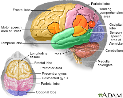
Growing brain tumors can place pressure on nearby parts of the brain. Brain swelling due to these tumors also causes increased pressure within the skull.
Brain tumors that spread are classified based on the location of the tumor in the brain, the type of tissue involved, and the original location of the tumor.
Metastatic brain tumors occur in about one fourth (25%) of all cancers that spread through the body. They are much more common than primary brain tumors (tumors that start in the brain).
Symptoms
Symptoms may include any of the following:
- Decreased coordination, clumsiness, falls
- General ill feeling or fatigue
- Headache, new or more severe than usual
- Memory loss, poor judgment, difficulty solving problems
- Numbness, tingling, pain, and other changes in sensation
- Personality changes
- Rapid emotional changes or strange behaviors
- Seizures that are new
- Problems with speech
- Vision changes, double vision, decreased vision
-
Vomiting, with or without nausea
- Weakness of a body area
Specific symptoms vary. Common symptoms of most types of metastatic brain tumors are caused by increased pressure in the brain.
Exams and Tests
An exam can show brain and nervous system changes based on where the tumor is in the brain. Signs of increased pressure in the skull are also common. Some tumors may not show signs until they are very large. Then, they can cause a very quick decline in nervous system function.
The original (primary) tumor may be found by examining tumor tissues from the brain.
Tests may include:
- Mammogram, CT scans of the chest, abdomen, and pelvis to find the original tumor site
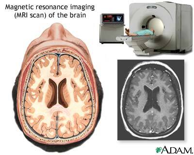
- CT scan or MRI of the brain to confirm the diagnosis and identify the tumor location (MRI is usually more sensitive for finding tumors in the brain)
- Examination of tissue removed from the tumor during surgery or CT scan- or MRI-guided biopsy to confirm the type of tumor
- Lumbar puncture (spinal tap)
Treatment
Treatment depends on:
- The size and type of the tumor
- Location in the body from where it spread
- The person's general health
The goals of treatment may be to relieve symptoms, improve functioning, or provide comfort.
Whole brain radiation therapy (WBRT) is often used to treat tumors that have spread to the brain, especially if there are many tumors, and surgery is not a good option.
Surgery may be used when there is a single tumor and the cancer has not spread to other parts of the body. Some tumors may be completely removed. Tumors that are deep or that extend into brain tissue may be reduced in size (debulked).
Surgery may reduce pressure and relieve symptoms in cases when the tumor cannot be removed.
Chemotherapy for metastatic brain tumors is usually not as helpful as surgery or radiation. Some types of tumors, though, do respond to chemotherapy.
Stereotactic radiosurgery (SRS) may also be used. This form of radiation therapy focuses high-power x-rays on a small area of the brain. It is used when there are only a few metastatic tumors.
Medicines for brain tumor symptoms include:
- Anticonvulsants such as phenytoin or levetiracetam to reduce or prevent seizures
- Corticosteroids such as dexamethasone to reduce brain swelling
- Pain medicines
When the cancer has spread, treatment may focus on relieving pain and other symptoms. This is called palliative or supportive care.
Comfort measures, safety measures, physical therapy, occupational therapy, and other treatments may improve the patient's quality of life. Some people may want to seek legal advice to help them create an advance directive and power of attorney for health care.
Support Groups
You can ease the stress of illness by joining a cancer support group. Sharing with others who have common experiences and problems can help you not feel alone.
Outlook (Prognosis)
For many people with metastatic brain tumors, the cancer is not curable. It will eventually spread to other areas of the body. Prognosis depends on the type of tumor and how it responds to treatment.
Possible Complications
Health problems that may result include:
- Brain herniation (fatal)
- Loss of ability to function or care for self
- Loss of ability to interact
- Permanent, severe loss of nervous system function that gets worse over time
When to Contact a Medical Professional
Call your health care provider if you develop a persistent headache that is new or different for you.
Call your provider or go to the emergency room if you or someone you know suddenly becomes sluggish or has vision changes, or speech impairment, or has seizures that are new or different.
References
Dorsey JF, Salinas RD, Dang M, et al. Cancer of the central nervous system. In: Niederhuber JE, Armitage JO, Kastan MB, Doroshow JH, Tepper JE, eds. Abeloff's Clinical Oncology. 6th ed. Philadelphia, PA: Elsevier; 2020:chap 63.
Elder JB, Nahed BV, Linskey ME, Olson JJ. Congress of Neurological Surgeons systematic review and evidence-based guidelines on the role of emerging and investigational therapies for the treatment of adults with metastatic brain tumors. Neurosurgery. 2019;84(3):E201-E203. PMID: 30629215 pubmed.ncbi.nlm.nih.gov/30629215/.
National Cancer Institute website. Adult central nervous system tumors treatment (PDQ) - health professional version. www.cancer.gov/types/brain/hp/adult-brain-treatment-pdq. Updated January 18, 2022. Accessed June 8, 2022.
Olson JJ, Kalkanis SN, Ryken TC. Congress of Neurological Surgeons Systematic Review and Evidence-Based Guidelines for the treatment of adults with metastatic brain tumors: executive summary. Neurosurgery. 2019;84(3):550-552. PMID: 30629218 pubmed.ncbi.nlm.nih.gov/30629218/.
Traylor JI, Rao G. Metastatic brain tumors. In: Winn HR, ed. Youmans and Winn Neurological Surgery. 8th ed. Philadelphia, PA: Elsevier; 2023:chap 169.