UF Health researchers use AI to improve brain tumor analysis

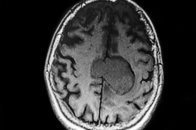
Five centimeters of a meningioma tumor is shown in this brain MRI. (Getty Image)
University of Florida Health researchers have deployed artificial intelligence to refine and accelerate the evaluation of a common type of brain tumor.
The findings involve analyzing certain chemical characteristics of meningioma tumors and then applying machine learning, a type of artificial intelligence. Those techniques help to identify the most relevant tumor features that doctors use to assign meningioma grades.
Correctly assessing a meningioma tumor is crucial because it drives a patient’s treatment plan: Slow-growing, less-threatening grade I tumors are removed and the patient gets follow-up monitoring. More aggressive, grade III tumors are typically removed and the site is treated with radiation. Grade II tumors pose more of a dilemma for physicians.
“Grade II tumors are the gray zone. Do we take it out and watch to see if it comes back? Or do we also irradiate the area with the idea of preventing a recurrence?” said study co-author Jesse L. Kresak, M.D., a clinical associate professor in the UF College of Medicine’s department of pathology, immunology and laboratory medicine. The findings were published recently in the Journal of the American Society for Mass Spectrometry.
The researchers analyzed 85 meningioma samples, focusing on comparing low-grade and high-grade tumors and identifying the biological markers that make them benign or malignant. To begin their study, the researchers obtained chemical profiles of the tumors’ fats and small molecules. That allowed them to better characterize the differences between grades of tumors and distinguish potential biomarkers that are helpful for diagnosis.
Using AI wasn’t always part of the researchers’ plan. The original idea was to analyze all of the byproducts of metabolism within tumor cells — a type of chemical fingerprint to help distinguish benign and malignant tumors.
That’s when Hoda Safari Yazd, Ph.D., who was a third-year graduate student at the time, proposed using machine learning.
“After talking about it, we knew that machine learning could be a good opportunity to find things that we wouldn’t be able to find ourselves,” said Timothy J. Garrett, Ph.D., a co-author of the paper, an associate professor in the College of Medicine’s department of pathology, immunology and laboratory medicine and a UF Health Cancer Center member.
This is how machine learning accelerates and improves the accuracy of the process: Kresak said she typically takes about 10 minutes and evaluates 20 data points when diagnosing a meningioma tumor. The machine learning model assessed nearly 17,000 data points in less than a second.
One of the machine-learning models also classified the grades of tumors with 87% initial accuracy, the researchers found. That figure can potentially be improved as more samples are analyzed.
While pathologists are skilled at diagnosing and grading brain tumors, Kresak said the AI and analytic techniques they studied can be helpful, powerful tools because tumors are sometimes reclassified based on their genetic makeup. The techniques used in the research would require federal regulators’ approval for use in diagnosis and treatment. Still, Kresak said assessing tumors on a metabolic level and using AI to analyze the data is a significant breakthrough.
“We are further understanding different tumors by using these tools. It’s a way to help us get the right treatment for our patients,” Kresak said.
Funding for the research was provided by the department of pathology, immunology and laboratory medicine, with scientific support from UF’s Southeast Center for Integrated Metabolomics.
About the author
