3D Printing of Replacement Talus Bone Potential Boon for Patients

The talus bone, all of two-inches long in adults, packs outsized influence in the mechanics of the foot and ankle.
It’s among the human bones with the highest percentage of its surface area covered by cartilage, more than 90%, as the talus forms joints with three bones — the tibia, calcaneus and navicular. The talus is the hub ensuring free and healthy movement of the foot and ankle.
Not surprisingly, an injury to the talus can be especially debilitating to a patient, with traditional surgery that, while beneficial, can come with significant trade-offs.
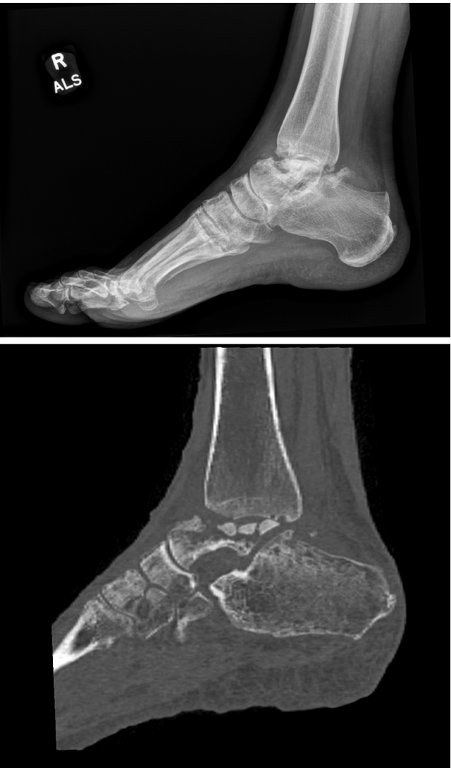
FIGURE 1A: Preoperative lateral X-ray (top) and sagittal CT scan (bottom) of the ankle demonstrating collapse of the talus, with resultant ankle and subtalar joint arthritis. (see Figure 1B below)
University of Florida Health recently joined the ranks of health systems nationally using a 3D printing technology that can build a new talus bone with a metal alloy to the exact specifications of a patient’s anatomy, allowing a faster recovery and providing a greater range of ankle motion after surgery, UF Health surgeons said.
“I really believe this is the future of talus surgery,” said UF Health orthopaedic surgeon R. James Toussaint, M.D., an associate professor and division chief in the UF College of Medicine’s foot and ankle service in the department of orthopaedic surgery and sports medicine.
“We have existing procedures that help address a diseased talus,” he added. “But their results may be unreliable especially if the talus has lost its blood supply. In addition, the procedures are labor intensive and time-consuming for the surgeon and patient. So, I think surgeons will increasingly embrace this 3D technology as they become more comfortable with it.”
The 3D talus implant is composed of a cobalt and chrome alloy, with specific dimensions customizable to a patient’s specific anatomy after a computerized tomography, or CT, scan of the patient’s normal foot and ankle.
Toussaint said the 3D technology for a talus replacement has been around for about a decade. But it has now matured to the point where large health systems like UF Health are beginning to increasingly use the technology.
“The process of 3D printing the metal alloy has improved significantly,” he said. “And the time it takes to create the artificial bone is substantially faster than before.”
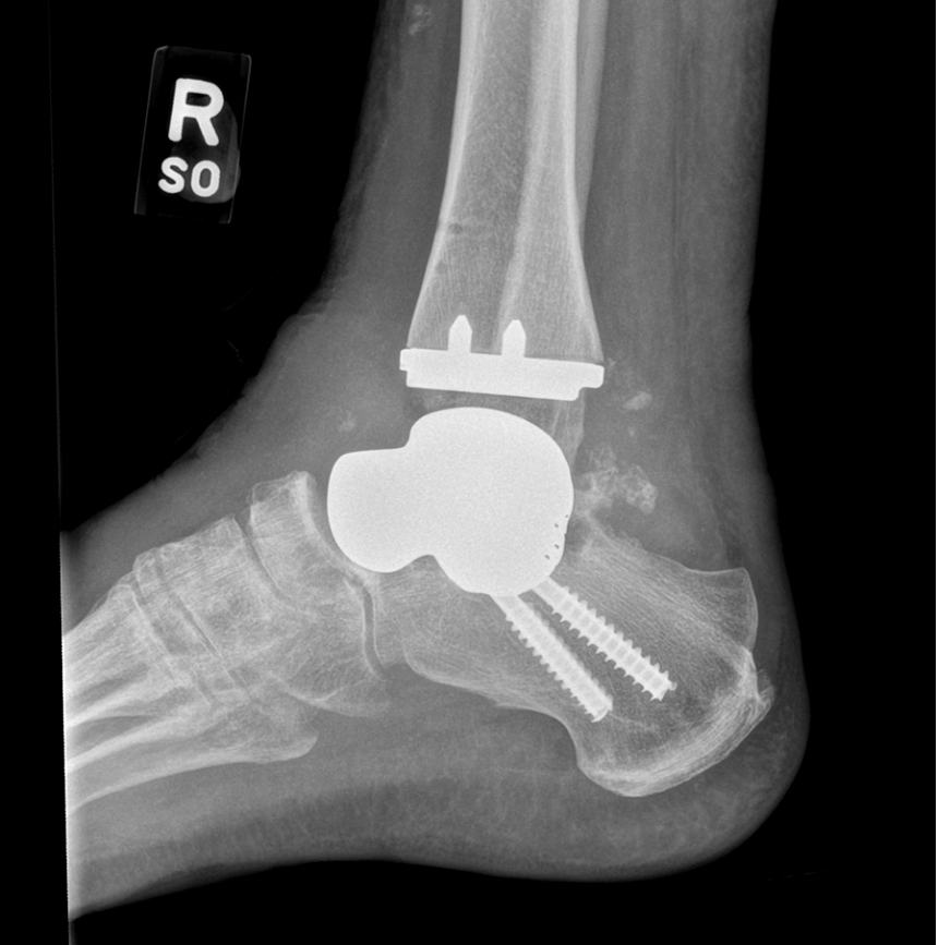
FIGURE 1B: Postoperative lateral X-ray of the ankle demonstrating a custom total talus replacement, ankle replacement and subtalar fusion.
A break of the talus can be devastating. The bone is well-protected in the foot, so it often requires severe trauma, such as a car accident or fall from a high place, to damage the bone. In fact, it is a relatively common trauma injury.
Indeed, golfer Tiger Woods broke his talus in a near-fatal car crash in 2021, an injury that, among others, imperils his career.
Talus injury can also occur in those people with vascular disease or have an alcohol-abuse disorder, Toussaint said.
In a severe injury, the talus’ tenuous blood supply is often disrupted, killing the bone and leading to a cascade of problems.
“What happens is that the bone collapses,” Toussaint said. “And as it collapses, it can affect the surrounding joints. They’re going to get arthritic.”
The gold standard treatment for arthritis from talus collapse has been bone fusion, but he noted it can be difficult to get the dead bone to fuse with adjoining live bone.
If the damaged talus causes arthritis at the ankle joint, then the joint can be replaced. But Toussaint states that is usually an option for older patients. Other cases, one may consider a fusion or an allograft using a cadaver bone that, however much the surgeon sculpts it, won’t be a precise match, which can further limit range of motion.
Additionally, he said, the surgeon also has the difficult task of finding a cadaver bone that matches the dimensions of the patient’s anatomy.
“A surgeon would have to find the exact match of a talus the same length and width of a patient’s own,” Toussaint noted. “You can imagine how daunting that can be.”
In some instances, patients have an option for amputation after failed surgery or pain that becomes difficult to bear.
The 3D-printed implant can more easily align with adjacent, healthy bone.
“Your options before 3D printing would have been to live with the pain,” Toussaint said. “You can inject pain medications. Or you could go through some pretty drastic surgery replacing dead bone with dead bone to achieve a fusion. And when you fuse those bones, the range of motion at that joint is eliminated.”
The implant is printed in North Carolina using proprietary technology developed by Restor3d, which ships the implant back to the surgeon within two to four weeks, Toussaint said.
“You then surgically remove the dead bone in the extremity and replace it instantly,” he said.
In a typical bone fusion, a patient has to stay off their foot for six weeks or more, until the surgeon confirms that a proper fusion is occurring.
“But with 3D printing the patient is walking within a week and a half or two weeks,” he said. “This offers patients a bastion of hope,” Toussaint said. “It’s giving them an option to preserve their function and potentially avoid a below-the-knee amputation.”
About the author
